

Ultrasound technology has brought about a revolution in the field of medical diagnostics. It offers non-invasive and real-time imaging capabilities that are incredibly valuable in healthcare settings.
Numerous specialty fields exist within the realm of ultrasound, and each one has its unique focus and applications. If you’re an ultrasound student or a seasoned professional looking for new opportunities, don’t worry. Understanding these specialty fields will help you find your niche and excel in your career.
So, let’s explore the specialty fields of ultrasound together, offering insights into their methodologies, applications, and career prospects. I’m sure you’ll find it fascinating and informative!
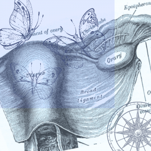
Obstetrics and Gynecology (OB/GYN) Ultrasound:
OB/GYN ultrasound is perhaps one of the most well-known specialty fields, focusing on imaging the female reproductive system during various stages of pregnancy and gynecological conditions. Here, sonographers are crucial in monitoring fetal development, detecting abnormalities, and assisting in prenatal care. This field offers a rewarding experience, allowing professionals to witness the miracle of life firsthand while providing essential healthcare support to expecting mothers.
There are various sub-specialties within the field of OB/GYN. Some sonographers specialize in working exclusively in fertility clinics, where they assist patients with procedures such as follicle monitoring, egg retrieval, and early ultrasounds.
In Maternal Fetal Medicine or Perinatal Sonography, perinatologists and sonographers with advanced imaging expertise work together to diagnose and treat maternal and fetal conditions. This sub-specialty also involves more advanced fetal heart views.
Cardiac Ultrasound (Echocardiography):
Cardiac ultrasound, or echocardiography, is a non-invasive medical procedure that involves capturing heart images to evaluate its structure and function. This diagnostic test is crucial in detecting and monitoring cardiovascular diseases, assessing the function of heart valves, and examining blood flow patterns.
Sonographers specializing in echocardiography must also deeply understand cardiac anatomy and physiology to properly interpret the images and identify any abnormalities or irregularities.
Cardiac ultrasound can be performed from various angles and planes, depending on the information needed and the condition being evaluated. The most common type of echocardiography is transthoracic echocardiography, where the transducer is placed on the chest wall to capture heart images. Other types include transesophageal echocardiography, where the transducer is inserted through the esophagus, and stress echocardiography, where the heart is evaluated before and after physical exercise.
Cardiac sonographers work closely with cardiologists and can further sub-specialize into pediatric, congenital, and fetal echocardiography.
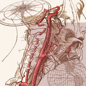 Vascular Ultrasound:
Vascular ultrasound is a non-invasive imaging technique that uses high-frequency sound waves to capture images of blood vessels throughout the body. This imaging modality is highly effective in diagnosing a range of vascular conditions, including deep vein thrombosis, venous reflux, peripheral artery disease, and aneurysms.
One of the key advantages of vascular ultrasound is that it allows technologists to visualize blood flow patterns within the vessels, which can help detect abnormalities indicative of underlying vascular diseases. Using Doppler ultrasound techniques, sonographers can measure the speed and direction of blood flow, providing valuable information about the health of the blood vessels.
Vascular ultrasound is particularly important in assessing the risk of stroke, heart attack, and other cardiovascular events. Medical professionals can prevent these serious health complications by detecting changes in blood flow patterns, blockages, or other abnormalities. Vascular sonographers work closely with vascular surgeons.
Musculoskeletal Ultrasound:
Musculoskeletal ultrasound is becoming increasingly popular for assessing injuries and conditions affecting the musculoskeletal system. This includes muscles, tendons, ligaments, and joints. Unlike traditional imaging modalities such as X-rays and MRI, musculoskeletal ultrasound provides real-time visualization of soft tissue structures and dynamic movement. It allows sports medicine practitioners, orthopedic surgeons, and rheumatologists to accurately diagnose and monitor musculoskeletal conditions.
Musculoskeletal ultrasound is particularly useful for identifying injuries and conditions that may not be visible on X-rays or MRI scans. It can detect small tears in muscles and tendons and inflammation in joints and soft tissue structures. This diagnostic tool can also guide interventions, such as injections and aspirations, and monitor treatment outcomes.
As a newer use of ultrasound, there is a lot of interest in MSK, but it is a challenging, evolving field. Becoming comfortable with assessing MSK anatomy requires a lot of time and expertise. MSK ultrasound is performed by sonographers and radiologists working together and learning the specialty.
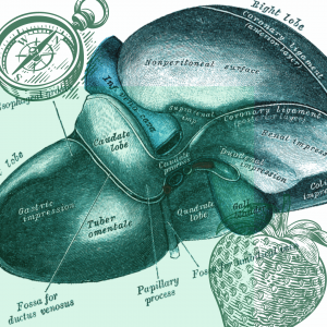 Abdominal Ultrasound:
Abdominal ultrasound is a type of diagnostic medical imaging involving sound waves to create images of the organs and structures within the abdominal cavity, including the liver, kidneys, pancreas, gallbladder, and spleen. In addition to these larger organs, abdominal ultrasound may include imaging of “small parts,” such as the thyroid, scrotum, and superficial lumps and bumps.
Abdominal ultrasound can provide valuable information about the health of organs and help diagnose a wide range of conditions. For example, it can detect gallstones in the gallbladder, liver cirrhosis, renal cysts, and abdominal masses. It can also be used to guide biopsies or other medical procedures.
Ultrasound techs specializing in abdominal ultrasound must be highly skilled and proficient in recognizing normal anatomy and identifying pathological changes that may indicate disease processes. There are over 300 diseases that can be diagnosed in the abdominal and small parts organs by ultrasound.
Pediatric Ultrasound:
Pediatric ultrasound is a specialized diagnostic medical imaging technique used to examine infants and children. It addresses the unique challenges posed by pediatric anatomy and physiology. Unlike adults, children’s organs and tissues are still developing, which can make interpreting ultrasound images more complex. As such, pediatric ultrasound technicians require specialized training and skills to perform this type of imaging.
Pediatric ultrasound is used to diagnose various medical conditions in children, including congenital anomalies, pediatric urological conditions, and abdominal disorders. Some of the specific conditions that can be diagnosed using this technique include appendicitis, kidney stones, bladder tumors, and liver disease. Ultrasound of the postnatal brain, hips, and spine is also unique to pediatric sonography.
One of the key challenges in pediatric ultrasound is ensuring the comfort and cooperation of young patients during the imaging procedure. This requires a high level of patience, empathy, and specialized skills. Pediatric ultrasound techs must communicate effectively with children and their parents and help alleviate any fears or concerns they may have. They must also be able to adapt their approach to imaging based on the child’s age, size, and medical condition.
 Breast Ultrasound:
Breast ultrasound plays a crucial role in diagnosing and evaluating breast abnormalities detected on mammography or clinical examination. It allows healthcare professionals to thoroughly examine the breast for any signs of cancer or other abnormalities.
The ultrasound is used to help characterize breast masses, cysts, and other lesions. It can also be used to guide biopsies, which is removing tissue for further examination under a microscope. Additionally, breast ultrasound is used to monitor patients with a history of breast cancer, as well as those who are at high risk of developing the disease.
Sonographers who specialize in breast ultrasound work closely with radiologists and breast surgeons to provide comprehensive breast imaging services. They play an important role in the early detection of breast cancer, which is essential for successful treatment.
Career Opportunities and Advancement:
The field of ultrasound is vast and offers numerous areas of specialization, each with its unique set of challenges and rewards.
It is not uncommon for sonographers to specialize in more than one modality. In fact, the main three branches: heart, vascular, and general (OB/GYN & Abdominal) have plenty of overlap. Many sonographers choose to learn more specialties to advance their career.
Becoming a sonographer usually starts with a college program focused on one of the big three (some programs do more than one). The educational program will include bookwork, practice scanning, and an internship.
To show competency in the specialization, a national credential exam is required. Credentialing organizations and sonography credentials offered include:
- ARDMS (American Registry for Diagnostic Medical Sonography)
- Abdomen, OB/GYN, Breast, Fetal Echocardiography, Pediatric Sonography, Adult Echocardiography, Pediatric Echocardiography, Vascular Technology, and Musculoskeletal Sonography
- CCI (Cardiovascular Credentialing International)
- Cardiac Sonography, Advanced Cardiac Sonography, Congenital Cardiac Sonography, Cardiac Sonography, Phlebology Sonography, and Vascular Sonography.
- ARRT (American Registry of Radiologic Technologists)
- General Sonography (includes abdominal and OB/GYN), Breast Sonography, and Vascular Sonography
With the increasing demand for sonographers, many job opportunities exist in various medical settings, including hospitals, clinics, imaging centers, and private practices. The demand for skilled techs is expected to grow as the healthcare industry expands and ultrasound imaging becomes more widely used for diagnostic and therapeutic purposes.
Pursuing a career in ultrasound specialization is an excellent choice for those who are passionate about the healthcare industry and want to positively impact people’s lives. With the right training, skills, and experience, professionals in this field can look forward to a rewarding career.
Finding your niche in the world of ultrasound involves exploring the diverse specialty fields and identifying the areas that resonate with your interests, skills, and career aspirations. Whether you’re drawn to the miracle of pregnancy, the intricacies of cardiac anatomy, or the dynamic nature of musculoskeletal imaging, there’s a specialty field waiting for you to explore. By gaining expertise in your chosen area and staying abreast of advancements in ultrasound technology, you can carve out a rewarding and fulfilling career path in this dynamic field of medical imaging.
Recent Comments